Aldehyde oxidase and xanthine dehydrogenase, a/b hammerhead domain
| Aldehyde oxidase and xanthine dehydrogenase, a/b hammerhead domain | |||||||
|---|---|---|---|---|---|---|---|
| Identifiers | |||||||
| Symbol | Ald_Xan_dh_C | ||||||
| Pfam | PF01315 | ||||||
| InterPro | IPR000674 | ||||||
| SCOP2 | 1alo / SCOPe / SUPFAM | ||||||
| |||||||
The aldehyde oxidase and xanthine dehydrogenase, α/β hammerhead domain is an evolutionary conserved protein domain. Both aldehyde oxidase (AOX) and xanthine dehydrogenase (XDH) contain this domain, along with other enzymes of the xanthine oxidase family.[1] The domain is a part of the molybdo-flavoenzyme family, and its shape contributes to the organization of the electron transfer occurring during the aldehyde oxidase and xanthine dehydrogenase catalytic cycle.[2]
Structure
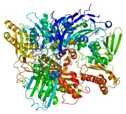
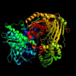
The α/β hammerhead domain is found in aldehyde oxidase and xanthine dehydrogenase. This domain has an α/β protein fold which are made up of alpha helices and beta sheets that are configured in a hammerhead shape.[3] They serve to help with the overall organization for the substrates within the active site, and stabilize the catalytic cofactors during electron transfer.[1] The catalytic cofactors active during electron transfer are molybdenum cofactor (MoCo), two [2Fe-2S] clusters, and flavin adenine dinucleotide (FAD). [1] MoCo is where the substrate binds and is oxidized, passing electrons to the molybdenum atom. The electrons then move through the two [2Fe-2S] clusters to the FAD, which is the final electron acceptor.
Function
Aldehyde oxidase (EC 1.2.3.1) catalyzes the conversion of an aldehyde in the presence of oxygen and water to an acid and hydrogen peroxide. The enzyme is a homodimer, and requires FAD, molybdenum and two 2FE-2S clusters as cofactors. Xanthine dehydrogenase (EC 1.1.1.204) catalyzes the hydrogenation of xanthine to urate, and also requires FAD, molybdenum and two 2FE-2S clusters as cofactors. This activity is often found in a bifunctional enzyme with xanthine oxidase (EC 1.1.3.22) activity too. The enzyme can be converted from the dehydrogenase form to the oxidase form irreversibly by proteolysis or reversibly through oxidation of sulfhydryl groups. After electrons reach FAD in the dehydrogenase form, FAD reduces NAD+ to NADH. In oxidase form the FAD gives the electrons to oxygen molecules, which ends up making superoxide or hydrogen peroxide. [1]
Species distribution
The aldehyde oxidase and xanthine dehydrogenase, a/b hammerhead domain is found in many prokaryotic and eukaryotic organisms, in enzymes of the AOX and XDH families.[1] Humans, plants, bacteria, and insects like fruit flies all have been identified molybdo-flavoenzymes, which have this domain.[4]
Clinical significance
For humans xanthine dehydrogenase play a major role when tissue perfusion is disrupted. During normal perfusion xanthine dehydrogenase usually functions in purine metabolism which doesn't produce many oxidative byproducts. However, in low oxygen or ischemic conditions, xanthine dehydrogenase is converted to xanthine oxidase by proteolysis.[1] Upon oxygen restoration, xanthine oxidase creates reactive oxygen species, especially superoxide and hydrogen peroxide.[5] Superoxide and hydrogen peroxide can lead to endothelial injury, impair microvascular function, and amplify inflammation. This contributes to the chain reaction that leads to ischemia-reperfusion damage occurring after events such as myocardial infarction.[5] Outside of perfusion related injury, mutations in the genes that encode for and xanthine dehydrogenase can lead to different disorders. Changes in the function of xanthine dehydrogenase can cause type I or type II xanthinuria, which can cause renal failure and kindey stones.[6]
See also
References
- ^ a b c d e f Garattini E, Mendel R, Romão MJ, Wright R, Terao M (May 2003). "Mammalian molybdo-flavoenzymes, an expanding family of proteins: structure, genetics, regulation, function and pathophysiology". The Biochemical Journal. 372 (Pt 1): 15–32. doi:10.1042/BJ20030121. PMC 1223366. PMID 12578558.
- ^ Harrison R (September 2002). "Structure and function of xanthine oxidoreductase: where are we now?". Free Radical Biology & Medicine. 33 (6): 774–797. Bibcode:2002FRBM...33..774H. doi:10.1016/S0891-5849(02)00956-5. PMID 12208366.
- ^ Bhat AS, Kinch LN, Grishin NV (November 2020). "β-Strand-mediated interactions of protein domains". Proteins. 88 (11): 1513–1527. doi:10.1002/prot.25970. PMC 8018532. PMID 32543729.
- ^ Amrani L, Primus J, Glatigny A, Arcangeli L, Scazzocchio C, Finnerty V (October 2000). "Comparison of the sequences of the Aspergillus nidulans hxB and Drosophila melanogaster ma-l genes with nifS from Azotobacter vinelandii suggests a mechanism for the insertion of the terminal sulphur atom in the molybdopterin cofactor". Molecular Microbiology. 38 (1): 114–125. doi:10.1046/j.1365-2958.2000.02119.x. PMID 11029694.
- ^ a b Granger DN, McCord JM, Parks DA, Hollwarth ME (January 1986). "Xanthine oxidase inhibitors attenuate ischemia-induced vascular permeability changes in the cat intestine". Gastroenterology. 90 (1): 80–84. doi:10.1016/0016-5085(86)90078-8. PMID 3753555.
- ^ "Hereditary xanthinuria: MedlinePlus Genetics". medlineplus.gov. Retrieved 2025-12-04.
Further reading
- Romão MJ, Archer M, Moura I, Moura JJ, LeGall J, Engh R, et al. (November 1995). "Crystal structure of the xanthine oxidase-related aldehyde oxido-reductase from D. gigas". Science. 270 (5239): 1170–1176. Bibcode:1995Sci...270.1170R. doi:10.1126/science.270.5239.1170. PMID 7502041. S2CID 34922450.
- Dobbek H, Gremer L, Meyer O, Huber R (August 1999). "Crystal structure and mechanism of CO dehydrogenase, a molybdo iron-sulfur flavoprotein containing S-selanylcysteine". Proceedings of the National Academy of Sciences of the United States of America. 96 (16): 8884–8889. Bibcode:1999PNAS...96.8884D. doi:10.1073/pnas.96.16.8884. PMC 17702. PMID 10430865.